

Elastography of the Shoulder: What Tendon Stiffness Tells Us About Pain, Healing and Recovery
Why Shoulder Imaging Needs to Go Beyond Structure
Shoulder pain is one of the most common musculoskeletal problems encountered in general practice. While MRI and conventional ultrasound are excellent at identifying structural abnormalities such as rotator cuff tears, they often fail to fully explain key clinical questions:
- Why do some patients experience significant pain despite minimal imaging changes?
- Why do some rotator cuff repairs fail while others heal successfully?
- Why does shoulder stiffness sometimes appear to protect against re-tear?
Recent research by our team has shown that the answers often lie not in structure alone, but in tendon quality and specifically, in tendon stiffness.
What Is Shoulder Elastography?
Shear-wave elastography is an advanced ultrasound technique that quantifies tissue stiffness in real time. It measures the speed at which ultrasound generated shear waves travel through tissue — with stiffer tissues transmitting waves faster.
Unlike conventional ultrasound, elastography provides:
- Objective, numerical measurements of stiffness (kPa or m/s)
- Colour-coded maps of tendon properties
- The ability to track tendon changes over time
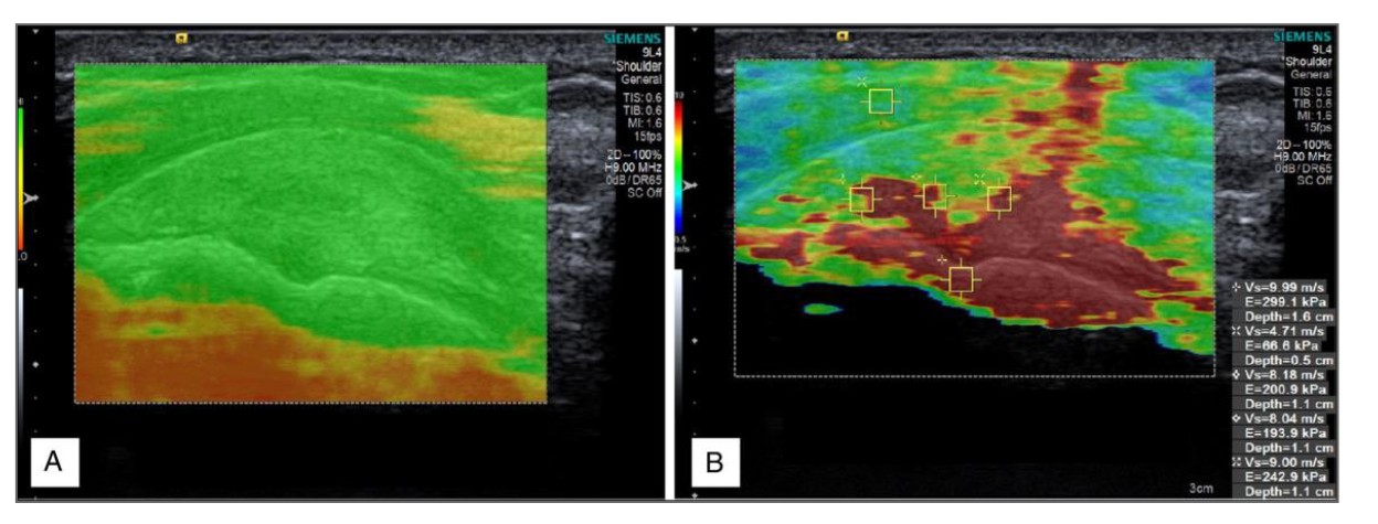
Figure 1. Standard B-mode ultrasound (A) compared with shear-wave elastography of the supraspinatus tendon (B).
Why Tendon Stiffness Matters in the Shoulder
The supraspinatus rotator cuff tendon plays a key role in shoulder function, holding the humeral head into the glenoid to provide a stable base for overhead function.
Elastography After Rotator Cuff Repair: Tracking Healing Over Time
Longitudinal studies conducted by Lisa Hackett during her PhD at UNSW have yielded the most comprehensive characterization to date of changes in supraspinatus tendon stiffness following rotator cuff repair.
Key findings include:
- Supraspinatus tendon stiffness increases by approximately 25% over 12 months
- The lateral tendon stiffens earlier than the medial tendon
- Older patients and those with larger tears demonstrate lower post-operative stiffness
- Stiffer tendons at 6–12 weeks are associated with better return to work and sport
- Elastography was shown to be more accurate than surgeon assessment alone in estimating tendon quality
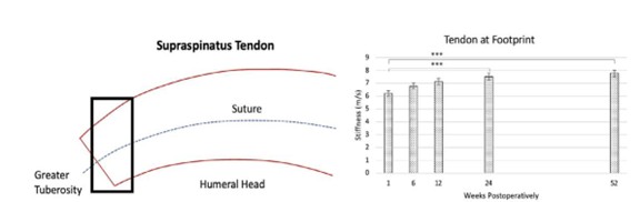
Figure 2. Temporal changes in the elastographic stiffness of supraspinatus tendon stiffness following rotator cuff repair.
Elastography and Frozen Shoulder: “There Is Gain With Pain”
Using shear-wave elastography, we have also demonstrated that patients with frozen shoulder exhibit marked thickening of the joint capsule, and a 60% increase in supraspinatus tendon stiffness compared with the unaffected side.
These findings provide a biological explanation for our previously documented clinical observation that patients with shoulder loss of motion tend to have lower rotator cuff re-tear rates after surgery.
Rather than stiffness being harmful, elastography suggests it reflects increased collagen deposition and a heightened healing response at the tendon–bone interface.
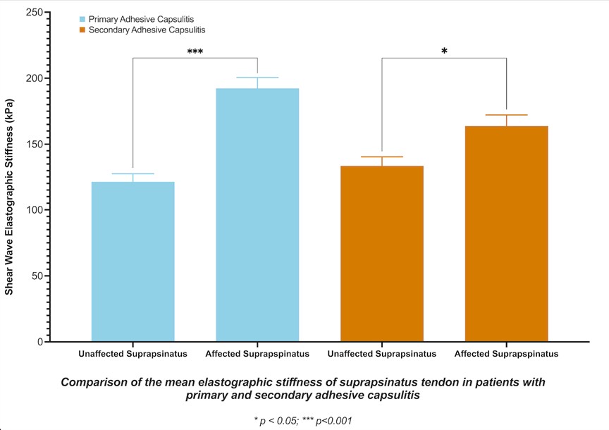
Figure 3. Elastography demonstrating increased supraspinatus tendon stiffness in adhesive capsulitis/Frozen Shoulder, particularly idiopathic/primary frozen shoulder.
Why This Matters for GPs and Referrers
For general practitioners, shoulder elastography can help explain persistent pain when MRI findings are inconclusive, identify patients at higher risk of delayed recovery or re-tear, assist with referral timing and shared decision-making, and provide an objective method to monitor healing and rehabilitation progress.
For patients, elastography offers a clearer answer to a common question:
“Is my tendon actually getting stronger?”
What Patients Experience
Shoulder elastography is non-invasive, painless, and radiation-free. It is performed alongside routine ultrasound and typically adds only a few minutes to the examination.
A Complement to Existing Imaging
Elastography does not replace MRI or conventional ultrasound. Instead, it adds functional insight, allowing clinicians to interpret structural findings in a biologically meaningful way.
Key Takeaway
Elastography of the shoulder provides objective insight into tendon health, healing, and prognosis that conventional imaging cannot. Research led by Professor George Murrell, with foundational work performed by Lisa Hackett and the Murrell UNSW research team, has established elastography as a powerful tool in understanding frozen shoulder, rotator cuff disease, and post-operative recovery.
Written by Professor George A. C. Murrell
Professor George Murrell is an Orthopaedic Surgeon who specialises in shoulder surgery, with interests in arthroscopic methods to repair and restore damaged ligaments and tendons.
About HealthShare
HealthShare is a leading digital health company. We are dedicated to improving health outcomes for patients through our innovative products for GPs, Specialists, Health organisations and more. To see how we can help you, visit HealthShare.